Dosya:Phytomyxea collage.jpg

Bu önizlemenin boyutu: 501 × 600 piksel. Diğer çözünürlükler: 200 × 240 piksel | 600 × 718 piksel.
Tam çözünürlük ((600 × 718 piksel, dosya boyutu: 201 KB, MIME tipi: image/jpeg))
Bu dosya Wikimedia Commons'ta bulunmaktadır. Dosyanın açıklaması aşağıda gösterilmiştir. Commons, serbest/özgür telifli medya dosyalarının bulundurulduğu depodur. Siz de yardım edebilirsiniz. |
Özet
| Açıklama |
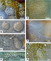
English: Morphology of resting spores from selected phytomyxids. Bar=10 μm.
|
| Tarih | |
| Kaynak | Neuhauser S., Kirchmair M., Bulman S., Bass D. (2014). "Cross-kingdom host shifts of phytomyxid parasites". BMC Evolutionary Biology 14 (33). DOI:10.1186/1471-2148-14-33. |
| Yazar | Sigrid Neuhauser, Martin Kirchmair, Simon Bulman and David Bass |
Lisanslama
Bu dosya, Creative Commons Atıf 2.0 Genel lisansı ile lisanslanmıştır
|
This file was published in a BioMed Central journal. Their website states that all of its research publications is published under the license which is identical to the Creative Commons Attribution 2.0 license (some non-research articles like reviews or editorials may require a subscription.)
To the uploader: You must provide a link (URL) to the original file or journal article.
|
Dosya geçmişi
Dosyanın herhangi bir zamandaki hâli için ilgili tarih/saat kısmına tıklayın.
| Tarih/Saat | Küçük resim | Boyutlar | Kullanıcı | Yorum | |
|---|---|---|---|---|---|
| güncel | 13.38, 24 Ocak 2016 | | 600 × 718 (201 KB) | Mithril | =={{int:filedesc}}== {{Information |Description= {{en|1=Morphology of resting spores from selected phytomyxids. Bar=10 μm: '''left column''': Plasmodiophorida, '''right column''': Phagomyxida. *'''A''': ''Sorosphaerula viticola'': hollow sporosori in... |
Dosya kullanımı
Bu görüntü dosyasına bağlantısı olan sayfalar:
Küresel dosya kullanımı
Aşağıdaki diğer vikiler bu dosyayı kullanır:
- ar.wikipedia.org üzerinde kullanımı
- arz.wikipedia.org üzerinde kullanımı
- en.wikipedia.org üzerinde kullanımı
- es.wikipedia.org üzerinde kullanımı
- hu.wikipedia.org üzerinde kullanımı
- ia.wikipedia.org üzerinde kullanımı
- ko.wikipedia.org üzerinde kullanımı
- pl.wikipedia.org üzerinde kullanımı
- ro.wikipedia.org üzerinde kullanımı
- ru.wikipedia.org üzerinde kullanımı
- www.wikidata.org üzerinde kullanımı